 In order to view your tissue sections under a microscope, they must be stained to make the cells visible. Most staining techniques use dyes that selectively stain tissues and cells in many different ways. We do this to help the pathologist identify the type of tissue present, and visualize changes in normal pathology or the presence of non-normal elements such as bacteria or fungus.
One of the most common routine stains is Hematoxylin and Eosin, often referred to as an H&E. The combination of a basic dye and an acidic dye allows the different elements of the tissue to be demonstrated with different colors. For example, nuclei stain blue and cytoplasm, muscle and red blood cells are a range of bright pink to red.
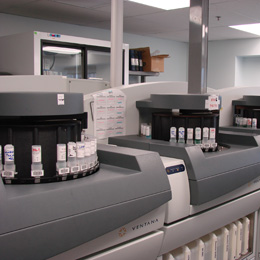
Automated instruments in our laboratory allow large volumes of slides to be stained without significant hands-on time for technical staff. The run time is from 20 – 40 minutes per slide batch. The stained slides are now ready for coverslipping.
Coverslipping
Racks of slides are moved from the stainer to the holding trough of a robotic coverslipper.
The purpose of the coverslipper is to apply a glass coverslip that completely covers the tissue section on the slide so that it's preserved. A drop of mounting media is dispensed onto the slide and the coverslip is placed on top. The mounting media acts as a glue to adhere the coverslip to the slide, and has an optical density close to that of glass so there is no distortion when viewing sections microscopically.
Quality control checks are performed before sending the slides to the pathologist. The slides prepared are documented in the LIS, and then delivered to the pathologist.
