By: Erin Vollick
It’s an electrifying step forward for cardiac research: a new method of maturing human heart cells that simulates their natural growth environment while applying electrical pulses to mimic a fetal heart rate.
The discovery, which offers cardiac researchers a fast and reliable way to create mature human cardiac patches in a range of sizes, was announced by University of Toronto researchers in the scientific journal Nature Methods.
“You cannot obtain human cardiomyocytes (heart cells) from human patients,” explained Milica Radisic, Canada Research Chair in Functional Cardiovascular Tissue Engineering and Associare Researcher, Toronto General Research Institute.
Because human heart cells—integral for studying the efficacy of cardiac drugs, for instance—do not naturally proliferate in large numbers, researchers have been using heart cells derived from reprogrammed human induced pluripotent stem cells (hiPSC’s), which tend to be too immature to use effectively in research or transplantation.
“The question is: if you want to test drugs or treat adult patients, do you want to use cells that look like and function like fetal cardiomyocytes?” asked Radisic, who is also Associate Professor with the Institute of Biomaterials and Biomedical Engineering and the Department of Chemical Engineering & Applied Chemistry.
In response to the challenge, Radisic and her team-- which includes Dr. Sara Nunes, a UHN scientist, and graduate student Jason Miklas-- created a ‘biowire' where human heart cells derived from stem cells are seeded along a silk suture typical to medical applications.
The suture allows the cells to grow along its length, close to their natural growth pattern.
Like a scene from Frankenstein, the cells are then treated to cycles of electric pulses which have been shown to stimulate the cells to increase in size, connect and beat like a real heart tissue.
But the key to successfully and rapidly maturing the cells turns out to be the way the pulses are applied.
Mimicking the conditions that occur naturally in fetal cardiac development—when the heart rate escalates before birth, the team ramped up the rate at which the cells were being stimulated, from zero to 180 and then 360 beats per minute.
“We found that pushing the cells to their limits over the course of a week derived the best effect,” said Radisic, who was named a “Top Innovator Under 35” by MIT Technology Review and more recently was awarded the Order of Ontario and the Young Engineers of Canada 2012 Achievement Award.
Grown on sutures that can be sewn directly into a patient, the biowires are designed to be fully transplantable. The use of biodegradable sutures, important in surgical patches that will remain in the body, is also a viable option.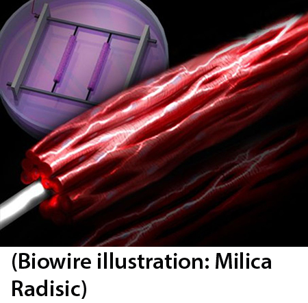
The research has practical implications for health care, said Miklas.
“With this discovery we can reduce costs on the health care system by creating more accurate drug screening.”
The development takes cardiac research one step closer to viable cardiac patches, said Nunes, a cardiac and a vascularization specialist.
“One of the greatest challenges of transplanting these patches is getting the cells to survive, and for that they need the blood vessels," Nunes said. "Our next challenge is to put the vascularization together with cardiac cells.”
Radisic, who calls the new method a “game changer,” is quick to point out just how far the field has come in a very short time.
“In 2006 science saw the first derivation of induced pluripotent stem cells from mice,” she said. “Now we can turn stem cells into cardiac cells and make relatively mature tissue from human samples, without ethical concerns.”
(The paper, "Biowire: a platform for maturation of human pluripotent stem cell–derived cardiomyocytes," can be found at
www.nature.com.)
Erin Vollick is a writer with U of T's Institute of Biomaterials and Biomedical Engineering.
