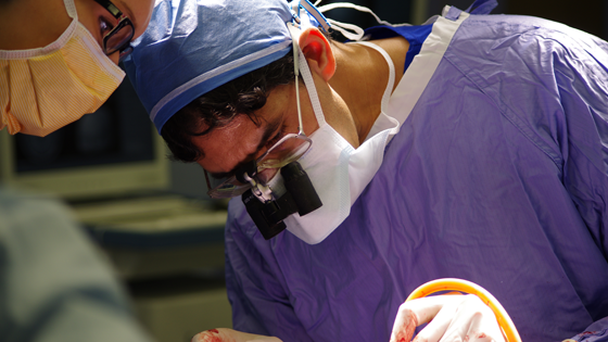
Dr. Taufik Valiante, neurosurgeon and co-director of KNC's Epilepsy Program, says that studying epilepsy has given experts the most insight into understanding the brain's functions. (Photo: UHN)
Did you know? A lot of what experts now know about the human brain is a result of studying one disease: epilepsy.
That's right – as doctors looked for ways to treat this often devastating neurological illness, they gained valuable insights to how the brain works and where its different functions lie.
Dr. Taufik Valiante, neurosurgeon and co-director of the epilepsy program at Toronto Western Hospital's Krembil Neuroscience Centre, discussed this interesting aspect of medical history with
UHNews. Check out what he had to say in the Q&A below:
UHNews: How did epilepsy come to play such a prominent role in our understanding of the human brain?
Dr. Valiante: One of Canada's most notable neurosurgeons, Dr. Wilder Penfield, is responsible for this. He pioneered a surgical treatment for severe epilepsy that came to be known as the "Montreal Procedure" – since he was based at McGill University in Montreal.
The procedure involved giving patients a local anesthetic so they could be kept awake during the operation. Penfield would then remove a part of the patient's skull to expose the brain. As he probed the brain with electricity, the patient could describe his/her reaction to Penfield which allowed him to identify the exact location of the seizure activity. Penfield would then remove the tissue causing seizures, and hopefully end the patient's seizures.
Some Canadians may recall the
Heritage Moment of this procedure where a patient describes the sensation of smelling burnt toast – a sign that seizure had started in a part of the brain involved in smell and memory – to Penfield.
At the time, more than half the patients treated with this new method were cured of seizures.
UHNews: What did experts learn about brain function through this technique?
Dr. Valiante: Penfield's technique gave him a lot of detail about the structure and function of the brain. This information allowed him to create maps of the sensory and motor sections of the brain, showing their connections to the various limbs and organs of the body. Because epilepsy can affect so many different parts of the brain, it is the only disease to give us such wide access to this organ.
These maps are still used today and have informed the whole field of Magnetic Resonance Imaging (MRI) research.
UHNews: Is this technique still in use today?
Dr. Valiante: Nowadays, we make use of electrical stimulation to map parts of the brain responsible for seizures – electrodes are implanted in the brain to diagnose where seizures are occurring.
The truest measure of what the brain does can be observed through its electrical activity – that's how different parts of the brain talk to each other and how it tells the rest of the body to do things: by emitting electrical signals. The brain obviously works very fast, emitting signals in thousandths of a second, so electrodes are the only thing that can give us a direct correlation between brain signal and brain function because they operate at the same speed.
When electrodes are implanted in the brain to monitor a patient's seizures, this allows us to observe whether stimulating an area of the brain blocks or enhances specific functions, such as speech, and lets us know that those functions are associated with that specific area of the brain. Mapping of this kind is more precise than what brain imaging can give us.
UHNews: What notable findings have experts gleaned from epilepsy patients?
Dr. Valiante: The most famous epilepsy patient was Henry Molaison, known as H.M. during the time he was studied from 1957 until his death in 2008. Molaison suffered from intractable epilepsy and had both of his hippocampi removed to treat his seizures. The hippocampus is a major component of the human brain located in the limbic system.
The removal of his hippocampi, unfortunately, caused Molaison to lose the ability to create new memories but helped medical experts discover that this area of the brain was involved in memory. His case was crucial in the development of theories that explain the link between brain function and memory, eventually establishing that the hippocampus plays an important role in the consolidation of information from short-term memory to long-term memory and spatial navigation.
Studying Molaison also led to the development of cognitive neuropsychology, a branch of psychology that aims to understand how the structure and function of the brain relates to specific psychological processes.
The brain tissue that was removed from Molaison's brain is still being studied.
UHNews: Is the information learned by studying epilepsy only applicable to brain research?
Dr. Valiante: Actually, no. The brain mapping that has occurred as a result of studying epilepsy has helped inform research in a number of different fields. For example, it has given new ideas to engineers on how to build better computers. Known as neuromorphic computing, this approach seeks to improve connections and processing of computer chips by following the principles of brain function.
Brain mapping has also given better insight into the mechanism of migraines – which has some features very similar to that of epilepsy since migraines are also caused by electrical disruption in the brain. And this research is also helping scientists develop algorithms for artificial intelligence to recognize things such as non-verbal communication.
Support epilepsy awareness
PURPLE DAY: It's time to break out your purple. Whether you sport a tie, don a dress or get decked out from head to toe, on Wednesday, March 26, wear purple to support Purple Day.
TWEET US YOUR PICS! Plus- Tweet pics of you in your purple to
@UHNews with #PurpleDay. We'll feature you in our Purple Day album on Facebook.
Related Links
Myths and facts about epilepsy
