You are looking at a strip of muscle, about the size of a grain of rice, suspended between two very fine wires. Despite its cylindrical shape, it is a miniature version of the human heart, and contracts and relaxes with the same hypnotic rhythm.
This feat was achieved by a team of researchers led by Drs.
Milica Radisic and
Peter Backx, Senior Scientists at the Toronto General Hospital Research Institute. Because their miniature heart closely mimics the cells found in a real heart, it may be a game changer in the study of heart disease and the search for therapeutic drugs.
The human heart has two types of chambers – atria and ventricles – that have different functions and are made of different kinds of cells. Because of this, atrial cells, which make up atria, and ventricular cells, which make up ventricles, respond to drugs differently.
While abnormal beating of the atria is the most common heart defect implicated in heart failure, up until recently, researchers had only been able to make ventricular tissues from human pluripotent stem cells.
The breakthrough platform engineered by Dr. Radisic and her colleagues, named Biowire II, is capable of growing both atrial and ventricular tissues. These atrio-ventricular Biowires have atrial cells on one end and ventricular cells on the other end, and have the same mechanical properties as real heart tissue.
"Biowires grown from the cells of healthy hearts are distinct from those grown from the cells of abnormal hearts," describes Dr. Radisic. "This enables us to model various heart conditions in the lab."
The Biowires also captured the effect of the drug ranolazine on the heart. Ranolazine is currently used to treat chest pain caused by insufficient blood supply to the heart.
With engineered tissues that resemble real hearts such as the Biowire II, researchers are one step closer to understanding the progression of heart disease and developing drugs that target specific heart cells without harming others.
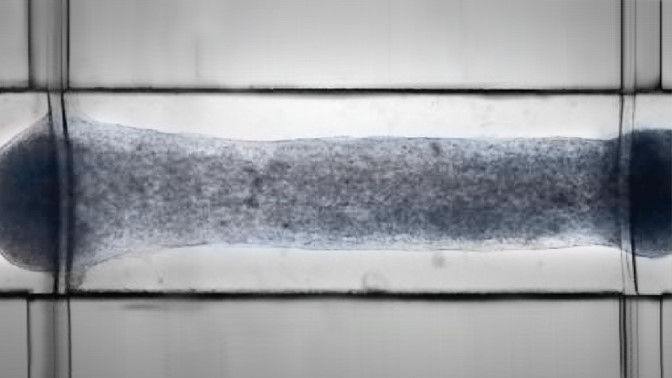
An atrio-ventricular Biowire under the microscope, suspended between two polymer wires. (Image: Courtesy Dr. Milica Radisic)
This work was supported by the Canadian Institutes of Health Research, the Natural Sciences and Engineering Research Council of Canada, the U.S. National Institutes of Health, the Ted Rogers Centre of Heart Research, and the Toronto General & Western Hospital Foundation. Dr. Brian Cox holds a Tier 2 Canada Research Chair (CRC) in Placental Development and Maternal-Fetal Health. Dr. Gordon M. Keller holds a Tier 1 CRC in Embryonic Stem Cell Biology. Dr. Peter H. Backx holds a Tier 1 CRC in Cardiovascular Biology. Dr. Milica Radisic holds a Tier 2 CRC in Functional Cardiovascular Tissue Engineering.
