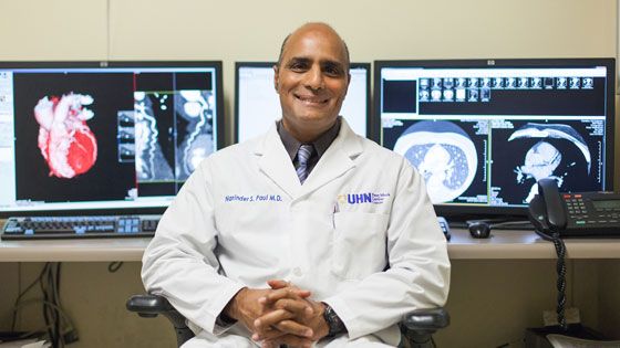
Dr. Narinder Paul, Division Chief, Cardiothoracic Imaging, Department of Medical Imaging, Peter Munk Cardiac Centre, Toronto General Hospital, suggests patients who are having their cardiac condition diagnosed ask a few key questions of their healthcare provider before undergoing a heart test. (Photo: UHN)
Nine in 10 Canadians have at least one risk factor for heart disease – but what is the best way to assess this? Better and early detection is central to addressing or even avoiding a potentially life-threatening cardiac event – in some cases, several years before it arrives.
Heart disease can occur for varying reasons, including: the narrowing of the coronary arteries (coronary atherosclerosis) or disease within the heart muscle (myocarditis, cardiomyopathy), the covering of the heart, the pericardium (pericarditis) or due to valve disease.
Tips on what to ask your doctor if you are asked to get a heart test:
- Why is this test necessary?
- What are the other options?
- In what ways will the results of the test impact my treatment plan?
- What kind of preparation does the test require?
- Should I refrain from taking any medications before the test?
While there is no single best test that can reliably evaluate all of these structures in the heart, here are some of the more common tests performed to assess heart disease:
-
12 lead Electrocardiogram (ECG): Tests the electrical activity of your heart.
How it works: As the heart squeezes and relaxes, it creates electrical patterns that can be recorded. Twelve leads are attached to the chest wall and to both arms and legs using small sticky pads. The heart pattern is recorded over a few heartbeats. The ECG can provide evidence of previous damage to the heart muscle or during an acute episode of chest pain. It can confirm whether there are any signs of future damage to the heart muscle. The test is useful as a baseline investigation but it is limited in providing a comprehensive assessment of coronary artery disease.
-
Treadmill Exercise Stress Test: Tests for coronary artery disease.
How it works: This test has been used for many decades to assess the likelihood of underlying coronary artery disease. It is similar to the 12 lead ECG but uses fewer leads and involves putting the heart under increasing stress. The patient walks on a treadmill using a set program that increases the speed and incline at regular intervals. Any significant narrowing of the coronary arteries will reproduce the patient's symptoms and result in changes in the ECG. This test is widely available. In patients who can exercise well, it provides an excellent assessment of heart health. However, the test is not useful in patients who have exercise limitation due to other medical conditions.
-
Trans-thoracic Echocardiography (TTE): Tests overall health of the heart.
How it works: TTE is essentially an ultrasound of the heart. The ultrasound machine is similar to the units used for performing diagnostic ultrasound elsewhere in the body. A small amount of ultrasound gel is placed on the chest wall to the left of the breastbone. A specialized ultrasound probe is gently applied to the chest wall and ultrasound waves are directed into the heart. TTE provides high-resolution images detailing the structure and function of the pericardium, the heart muscle and of the valves, in real-time. It also provides detailed information on the flow of blood through the heart chambers and into the aorta. Traditional TTE is very useful, safe, portable and can be performed at the bedside. In some patients TTE is limited by the shape or size of the chest wall, or by the lungs that envelope the heart as ultrasound cannot travel through air.
-
Trans-Esophageal echocardiography (TEE): A test that produces pictures of the heart.
How it works: TEE is performed when ultra-high resolution images of the heart are required. A small ultrasound probe is placed on the end of a fiber-optic endoscope that is gently guided through the mouth and into the esophagus. The scope is advanced until it sits just above the level of the diaphragm and is in contact with the left atrium through the wall of the esophagus.
-
Stress Echocardiography: Tests how well your heart and blood vessels are working.
How it works: Neither traditional TTE or TEE provide assessment of coronary artery disease. Stress echocardiography is TTE performed before and after the patient has exercised on a treadmill or on a stationary bicycle. If the patient cannot exercise then a drug is injected intravenously to simulate the effect of exercise. If there is significant coronary artery narrowing, the heart muscle that is supplied by this artery does not move in a normal way and this can be seen on the TTE. Stress echocardiography has the same limitations as conventional TTE.
-
Myocardial Perfusion Imaging (MPI): A non-invasive imaging test that shows how well blood flows through (perfuses) your heart muscle.
How it works: MPI requires the injection of a radioactive dye that circulates in the blood steam and is taken up into the heart muscle (myocardium). The radioisotope produces a special form of radiation detected by a gamma camera. Images of the heart muscle are taken from various angles during exercise and at rest. The radioisotope is not detected in areas with poor blood supply. If a normal uptake of radioisotope is seen at rest and stress, there is no significant coronary artery disease. A lack of radioisotope during stress and rest indicates permanent blockage in the blood supply to this area. A normal uptake at rest with abnormal uptake during stress indicates significant narrowing of the coronary arteries.
-
Cardiac Magnetic Resonance Imaging (MRI): An imaging method that uses powerful magnets and radio waves to create pictures of the heart.
How it works: Cardiac MRI uses magnetic waves to provide detailed images of the heart from all possible angles, providing detailed structural and functional images of the heart. MRI is also used to perform a detailed evaluation of the heart muscle including assessing swelling as a result of infection, infiltration by tumours and abnormal muscle growth due to certain heart disorders.
-
Perfusion Myocardial Magnetic Resonance Imaging (Perfusion MRI): A test for patients with known or suspected coronary artery disease.
How it works: This test can be performed at rest in patients who have a history of a prior heart attack to determine if coronary artery bypass surgery will be successful in reinvigorating the heart muscle. Cardiac MRI images are taken 15 minutes after injection of a dye that follows the blood flow into the heart muscle. The dye remains in areas of myocardial scar tissue. The test can also be performed during exercise or stress; the dye is injected during peak myocardial stress and at rest to evaluate areas of the myocardium that might have a severely compromised blood supply. MRI provides high resolution, detailed images of the heart and there is no radiation exposure.
-
Cardiac Computed Tomography (CCT): A test that uses an x-ray machine to take clear, detailed pictures of the heart.
How it works: Ongoing improvements in computed tomography (CT) scanners have resulted in ultrafast, high resolution imaging of the heart. The CT images of the heart and coronary arteries are synchronized with the heartbeat. Ideally, the heart rate needs to be steady at 60 beats per minute or less to minimize blurring of the coronary arteries due to motion.
-
Coronary Calcium Score (CCS): Tests the amount of hardening of the artery wall.
How it works: The CCS is used to assess patients who have coronary risk factors without any symptoms of coronary artery disease. CCS is performed without injection of a dye and the images are used to calculate the amount of calcium in the coronary arteries. Once calculated, the total amount of coronary calcium is compared to a database containing people of the same age and sex in order to assess the relative amount of calcified plaque. This allows patients to be ranked on a percentile chart and provide an assessment for the potential of future coronary events.
-
Coronary CT angiography (CCTA): An imaging test that looks at the arteries that supply your heart with blood.
How it works: CCTA is primarily used to evaluate the coronary arteries in patients who have risk factors for coronary artery disease and symptoms that are possibly due to heart disease but remain unclear. A dye containing iodine is injected and follows the blood supply. CT images are obtained as the dye passes through the coronary arteries. The patient must hold their breath for 1-8 seconds depending on the type of CT scanner. CCTA is the best non-invasive test for producing images of the coronary arteries and has a very high accuracy (more than 98 per cent) for detecting coronary artery disease. However, access to CCTA is limited due to expertise and a very irregular or very high heart rate may be challenging to produce clear images.
-
Catheter Coronary Angiography (CCA): Tests how blood flows through the arteries in your heart using a special dye and x-rays.
How it works: The best test for confirming the presence of significant coronary artery disease, CCA involves puncturing an artery in the groin; guiding a thin plastic tube (catheter) through the arterial system and into the coronary arteries. A dye containing iodine is injected into each of the coronary arteries and multiple X-ray images are taken at a high frame rate to capture the dye as it fills the coronary arteries. CCA is the most common imaging test performed if a patient presents with chest pain and the 12 lead ECG suggests severe coronary artery disease. Once the catheter is in position in the coronary artery, it may be possible to treat a seriously narrowed coronary artery by inserting a stent. However, CCA is invasive and a small number of patients develop a bruise at the puncture site.
