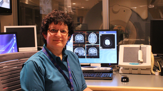
Dr. Karen Davis, pictured in one of the MRI tech rooms of TWH, uses imaging techniques to study how the human brain processes and reacts to pain. (Photo: UHN)
As scientists learn more about the brain, they have come to understand that its different regions are responsible for diverse things. The hippocampus is associated with memory. The amygdala is the centre for processing decisions and emotions. The prefrontal area regulates personality and emotional traits.
But when it comes to pain, the entire brain receives signals when something hurts, which is partly what makes it so difficult to treat.
"Pain is a distributed system where its signals are sent to many areas in the brain," says Dr. Karen Davis, Senior Scientist at Toronto Western Research Institute (TWRI) and an expert in pain research.
"The challenge of finding treatments for chronic pain is that it's hard to target something that is felt everywhere."
Related to this story:
Researchers' greater understanding of the brain has also led to more insight about chronic pain, and Dr. Davis is leading the charge.
Her research, which uses imaging techniques to study how the brain responds to pain, has shown that people process pain differently. Because of that, some may benefit more from some treatments than others.
"Seventy per cent of patients suffering from chronic pain have no benefit from the treatments currently available. This is an enormous number," Davis explains.
"I'm a fundamental scientist at the core; I like to understand how things work. Through a better understanding of how people's brains interpret and react to pain, my hope is that it will eventually change clinical practice in treating these patients for the better."
Pain is personal
Dr. Davis was the first scientist to publish research showing that pain - and
how we cope with it - is a very personal experience. Over the last decade, she has continued to investigate these findings with the underlying hypothesis that this variance in pain experience most likely shows differences in brain structure and function.
Being able to not only figure out but also draw a blueprint of these differing structures and how they operate will lead to individualized 'pain profiles.' This is a type of cheat sheet that could tell medical experts whether or not a particular patient would benefit from a pain treatment they are considering.
Dr. Davis'
most recent paper, published in the
Journal of Neuroscience this month, found that when it comes to brain regulation of pain, people generally fall into one of two groups: antinociceptive, where a person's brain system triggers pain modulation so that they experience less pain, and pronociceptive, which are individuals whose brains are very sensitive to incoming pain signals.
Moving forward
Now that Dr. Davis and her team have identified these differences, the next phase is to determine how rigid they are and whether a pronociceptive brain can become antinociceptive.
"It's becoming apparent that, when it comes to pain, everyone has their own personalized circuitry. But because there is widespread inter-connectiveness across vast regions of the brain, it's a bit like the block-stacking Jenga game – how do you tap into one area in this system without negatively affecting the brain as a whole?" says Dr. Davis. "With all the technology we have today, we should be able to figure out how this system works."
It's because of the advances made in technology over the last 25 years that Dr. Davis entered this area of research.
'Cartographer of the brain's pain system'
Trained as an electrophysiologist, she was hired at Toronto Western Hospital (TWH) mostly to do intraoperative mapping – measuring the electrical activity of neurons – to guide surgeons during procedures in the brain. Dr. Davis would also use the data from these procedures for her research, but the information it provided was limited to the brain areas being operated on.
"At the time, because these patients were already in need of care, we were only getting a small picture of what a sick brain looked like," she recalls. "We were unable to make comparisons to healthy brains because there was no existing tool we could use to study them."
But this didn't last for long.
Over the course of the 90s, magnetic resonance imaging (MRI), functional magnetic resonance imaging (fMRI), and positron emission tomography (PET) scans became more widely available and used for diagnostic testing. And because MRIs were non-invasive, it was possible for researchers to study the internal tissues and organs of healthy individuals.
It was the opportunity Dr. Davis needed to further her research about the brain.
"With the arrival of fMRI, we could now look at the whole brain in both healthy and sick patients," she says. "The research possibilities increased enormously because scientists could now ask questions beyond what could be answered studying one cell at a time."
Twenty years after first embarking on her quest as cartographer of the brain's pain system, Dr. Davis is confident medical experts are getting closer to realizing personal 'maps of pain' for their patients.
Knowing how a patient's brain processes pain will help them avoid needless procedures that may not alleviate their pain, and guide practitioners to treatments that will help – an approach now known as personalized precision medicine.
