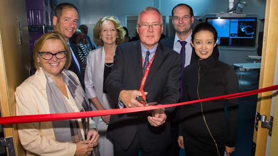
Delivering state-of-the-art care: UHN doctors and leaders cut the ribbon to mark the opening of two new treatment rooms. (UHN PhotoGraphics)
The Joint Department of Medical Imaging (JDMI) officially opened two new procedure rooms at Toronto Western Hospital (TWH), with biplane angiography systems that produce highly detailed views of the brain.
Using Philips AlluraClarity imaging technology, interventional neuro-radiologists now have the ability to perform a new range of treatments for conditions like stroke, aneurysms and vascular conditions in the brain.
"We now have specialized equipment that matches the world-leading expertise of our interventional neuro-radiologists," said Dr. Karl terBrugge, who helped spearhead the project as Head of Neuro-radiology at TWH.
'From paper to reality'
"With this new technology we are able to see the brain in greater detail and access spaces that we are not normally able to see, such as tiny blood vessels, and perform treatments with tools that are no thicker than the size of a strand of hair."
The collaboration and dedication of teams across UHN to renovate the space resulted in a "remarkable" turnaround time.
"In just one year, the plan was taken from paper to reality – giving doctors the ability to save lives, perform ground-breaking research and provide education across the globe," said Catherine Wang, Executive Director of JDMI.
Lorraine's story
Lorraine, one of the first patients treated with the new equipment, attended the ribbon cutting event to share her remarkable story of recovery after being diagnosed and treated by Dr. Timo Krings for a dural arteriovenous fistula of the cavernous sinus.
Simply put, there was an abnormal connection between an artery and vein in her brain – and left untreated, could have resulted in a brain hemorrhage.
Dr. Krings inserted catheters into both her arteries and veins and by combining preoperatively obtained cross-sectional data, he was able to find a pathway to the lesion that was not visible on conventional Angiography alone. Once in place he blocked the leakage with tiny platinum spirals leading to immediate resolution of Lorraine's symptoms.
Here's how many more patients will benefit from our new procedure rooms:
-
Stroke: New imaging equipment helps to identify and assess the size and direction of a blocked artery during acute stroke, allowing the neuro-radiologists to look at where the clot starts and stops to perform safer treatments.
-
Complex brain vascular malformations (abnormal connection of blood vessels): We are now able to fuse preoperative imaging (such as MR or CT) into the neuro-angiography procedure room to better define the tangled blood vessels and the surrounding brain.
-
Brain aneurysm: Continuous three dimensional image of brain aneurysms allow for more precise treatment. New software developed by our researchers in partnership with Philips allows us to predict treatment outcomes during the procedure using optical flow imaging.
"In interventional neuroradiology we are always striving to see inside the brain beyond the blood vessel," said Dr. Krings.
"This new equipment allows us to do what few other hospitals in Canada are able to do – combine MRI and CT in real time with the angiographical images, with the added ability to perform advanced research that will lead to the next generation of treatments."
